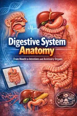

Free ebook on digestive system anatomy, covering the GI tract, liver, pancreas, gallbladder, vessels, nerves, and key landmarks.
Free ebook content
-

Digestive System Anatomy Map: Head-to-Toe Organization and Key Landmarks
+ Exercise: Which structure is considered an accessory digestive organ because it adds secretions into the GI tract rather than being part of the continuous hollow tube?
-
Foundations of the Gastrointestinal Wall: Layers, Plexuses, and Regional Specializations
+ Exercise: A patient has impaired peristalsis and abnormal sphincter relaxation, while mucosal secretion appears relatively normal. Which structure is most likely disrupted?
-
Peritoneum and Abdominal Compartments: Intraperitoneal, Retroperitoneal, and Mesenteries
+ Exercise: Which statement best distinguishes an intraperitoneal organ from a secondarily retroperitoneal organ?


-
Oral Cavity Anatomy for Digestion: Teeth, Tongue, Salivary Pathways, and Swallowing Entry
+ Exercise: During swallowing, which action helps prevent the bolus from entering the nasopharynx as it moves through the fauces into the oropharynx?
-
Pharynx and Esophagus: Conduits, Constrictions, and Sphincteric Regions
+ Exercise: Which statement best explains why the lower esophageal sphincter (LES) region helps prevent reflux?
-
Stomach Anatomy: Regions, Curvatures, Omenta Attachments, and Wall Features
+ Exercise: During a dissection, you want to orient the stomach by using its curvatures to predict peritoneal attachments. Which statement is correct?
-
Small Intestine Anatomy: Duodenum, Jejunum, Ileum, and Absorptive Surface Design
+ Exercise: Which set of features most reliably indicates a segment of ileum rather than jejunum?
-
Pancreas and Biliary Anatomy: Duct Systems and Duodenal Integration
+ Exercise: Which structure typically regulates the flow of both bile and pancreatic juice into the duodenum at the major duodenal papilla?
-
Liver Anatomy: Lobes, Porta Hepatis, Peritoneal Reflections, and Segment Orientation
+ Exercise: Which statement correctly describes the porta hepatis and what structures pass through it?
-
Gallbladder and Extrahepatic Bile Ducts: Storage Anatomy and Clinical Landmarks
+ Exercise: When tracing bile ducts from the gallbladder toward the duodenum, which identification best describes the duct formed at the junction of the cystic duct and the common hepatic duct?
-
Large Intestine Anatomy: Cecum, Appendix, Colon Segments, and Haustra Features
+ Exercise: When distinguishing large intestine from small intestine during dissection or imaging, which combination of external features most strongly supports that a segment is colon rather than small intestine?
-
Rectum and Anal Canal Anatomy: Pelvic Relations and Sphincter Complex
+ Exercise: Which statement best describes how the puborectalis contributes to continence at the anorectal junction?
-
GI Vasculature, Lymphatics, and Innervation: Regional Supply Patterns from Foregut to Hindgut
+ Exercise: A lesion is located in the distal one-third of the transverse colon near the splenic flexure. Which combination best predicts its primary arterial supply and typical venous drainage pathway?
-
Integrated Topography: Putting the Digestive Organs Together in Cross-Sections and Surface Projections
+ Exercise: On an axial abdominal image at the umbilical level, which pattern of digestive structures is most consistent with integrated topography?
About the free ebook
Digestive System Anatomy: From Mouth to Intestines and Accessory Organs
This free ebook provides a structured anatomy guide to the digestive system, following the route of food from the oral cavity through the pharynx, esophagus, stomach, small intestine, large intestine, rectum, and anal canal.
Understand the anatomy behind digestion
Learn how the gastrointestinal tract is organized, including its wall layers, enteric nerve plexuses, regional specializations, peritoneal relationships, mesenteries, and abdominal compartments. Clear anatomical landmarks support an integrated view of organ position and connection.
Explore the major digestive organs
- Oral cavity structures, teeth, tongue, salivary pathways, and swallowing entry
- Esophageal constrictions, stomach regions, curvatures, and omental attachments
- Duodenum, jejunum, ileum, and features that increase absorptive surface
- Cecum, appendix, colon, rectum, anal canal, and sphincter complex
Study the accessory organs and their connections
Examine the liver, gallbladder, pancreas, and biliary duct system, with attention to the porta hepatis, pancreatic ducts, bile flow, and duodenal integration. The ebook also connects digestive anatomy with arterial supply, venous drainage, lymphatics, and innervation patterns across the foregut, midgut, and hindgut.
Build a spatial understanding
Use cross-sectional and surface-projection perspectives to relate digestive organs to one another. This anatomy resource is useful for reviewing landmarks, pathways, tissue organization, and clinical orientation within the abdomen and pelvis.
What are the four layers of the gastrointestinal wall?
Most of the GI tract has mucosa, submucosa, muscularis externa, and serosa or adventitia.
Which digestive organs are retroperitoneal?
Key retroperitoneal structures include most of the duodenum, pancreas, and ascending and descending colon.
How do bile and pancreatic secretions enter the duodenum?
They travel through bile and pancreatic ducts and commonly enter at the major duodenal papilla.
This ebook includes:

14 content chapters

Digital certificate of course completion (Free)

Exercises to train your knowledge

100% free, from content to certificate
Ready to get started?

 recommended
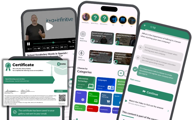
In the app you will also find...

Over 5,000 free courses
Programming, English, Digital Marketing and much more! Learn whatever you want, for free.

Study plan with AI
Our app's Artificial Intelligence can create a study schedule for the course you choose.

From zero to professional success
Improve your resume with our free Certificate and then use our Artificial Intelligence to find your dream job.
You can also use the QR Code or the links below.