Duration of the online course: 2 hours and 49 minutes
3
(4)
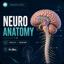
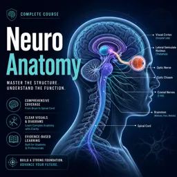
Strengthen your neuroanatomy skills with this free online course—master cranial nerves, thalamus, and brain vessels through clear videos and quick quizzes.
In this free course, learn about
Dural venous sinuses: anatomy and role in venous drainage and CSF absorption
Superficial brain veins: locations and function in cortical venous drainage
Spinal cord blood supply/drainage: key arteries, veins, and vascular territories
Spinal meninges & nerve roots: layers, spaces, and protective/support roles
Pia mater: structure and primary function in covering/nourishing the CNS
Thalamic nuclei: major divisions and relay functions (“traffic control” to cortex)
Brain section anatomy: identify structures in horizontal/coronal views incl. ventricles
Cerebellar nuclei/cerebellum: roles in coordination, balance, and motor learning
Cranial nerves overview: list, core functions, and key clinical associations
CN IX glossopharyngeal: branches and sensory supply incl. pharyngotympanic tube
CN VIII vestibulocochlear: anatomy/function and symptoms of damage (hearing/vertigo)
Eye movement nerves (III, IV, VI) & V1: functions; lateral rectus innervation by CN VI
CN XI accessory & CN XII hypoglossal: motor targets and main actions (SCM/trapezius, tongue)
About the free online course
Understanding neuroanatomy can feel overwhelming when structures, pathways, and clinical effects blur together. This free online course helps you build a clear mental map of the nervous system by focusing on the parts that students and health professionals use most: protective coverings, vascular routes, key brain regions, and the cranial nerves that connect brain to body.
Through concise lessons and targeted practice questions, you will connect anatomy with function in a way that supports exams and real-world reasoning. You will follow how blood drains from the brain through dural venous sinuses, how vessels support the spinal cord, and how the meninges—especially the pia mater—relate to the central nervous system. These foundations make it easier to interpret what is happening when neurological function is affected.
The course also sharpens your ability to localize and explain findings by exploring major neural structures and pathways. You will study thalamic nuclei and the thalamus as a relay center that organizes sensory and motor signals headed to the cerebral cortex. You will strengthen spatial understanding with brain sections, learning how to orient yourself on horizontal and coronal views and recognize ventricular landmarks that guide navigation through the brain.
Cranial nerves receive special attention to support both anatomy learning and clinical correlation. You will clarify the roles of nerves involved in sensation, swallowing, eye movement, hearing and balance, neck and shoulder control, and tongue movement. In parallel, you will link nuclei and brainstem locations to primary functions, so the names and pathways become meaningful rather than memorized facts.
By the end, you should feel more confident describing core neuroanatomy, explaining what structures do, and answering typical exam-style questions about functions and deficits. Whether you are in a health program or refreshing anatomy for practice, this course offers a structured, approachable way to study and retain neuroanatomy online.
Course content
Video class: Dural venous sinuses (preview) - Human Anatomy | Kenhub02m
Exercise: What is the primary function of the dural venous sinuses in the brain?
Video class: Blood Vessels of the Spinal Cord (preview) - Human Anatomy | Kenhub02m
Exercise: Which of the following structures are involved in the vascular supply and drainage of the spinal cord?
Video class: Glossopharyngeal Nerve Overview in 5 minutes - Human Anatomy | Kenhub05m
Exercise: Which of the following branches of the glossopharyngeal nerve is responsible for transmitting sensory information from the pharyngotympanic tube?
Video class: What happens when the vestibulocochlear nerve is damaged? - Human Anatomy | Kenhub03m
Exercise: What symptoms are commonly associated with damage to the vestibulocochlear nerve?
Video class: Thalamic nuclei: anatomy and functions (preview) - Human Neuroanatomy | Kenhub03m
Exercise: What role does the thalamus play in the human brain, similar to a function performed in air traffic management?
Exercise: What role does the thalamus play in the brain, as compared to air traffic controllers?
Video class: The Pia Mater (Meninges of the Brain) - Human Anatomy | Kenhub04m
Exercise: Which of the following describes the primary function of the pia mater in the central nervous system?
Video class: All About the Ophthalmic Nerve (preview) - Human Anatomy | Kenhub02m
Exercise: Which of the following is the first branch of the trigeminal nerve also known as?
Video class: Oculomotor, Trochlear and Abducens Nerves (preview) - Human Anatomy | Kenhub02m
Exercise: Which cranial nerve is primarily responsible for innervating the lateral rectus muscle that abducts the eye?
Video class: What is the Accessory Nerve? (preview) - Human Anatomy | Kenhub02m
Exercise: Which cranial nerve is known for innervating the sternocleidomastoid and trapezius muscles, and is displayed prominently in green in the tutorial image from an anterior view?
Video class: Vestibulocochlear nerve: anatomy and function (preview) - Human Neuroanatomy | Kenhub03m
Video class: Hypoglossal nerve: course and branches (preview) - Human Neuroanatomy | Kenhub03m
Exercise: What is the main function of the hypoglossal nerve?
Video class: Cranial nerve nuclei: locations and functions (preview) - Human Neuroanatomy | Kenhub03m
Exercise: In which part of the brainstem are the cranial nerve nuclei primarily located, and what are their primary functions?
Video class: Cranial nerves: list and functions (preview) - Human Anatomy | Kenhub03m
Exercise: Which cranial nerve is responsible for transmitting visual impulses from the retina to the brain?
Video class: Cerebellar nuclei: anatomy and functions (preview) - Human Neuroanatomy | Kenhub03m
Exercise: Which of the following is a primary function of the cerebellum?
Video class: Meninges of the Brain Overview (preview) - Human Anatomy | Kenhub03m
Exercise: Which is the strongest membrane covering the brain, also known for forming the dural venous sinuses?
Video class: Maxillary nerve: Branches, course (preview) - Human Anatomy | Kenhub03m
Exercise: What is the primary function of the maxillary nerve?
Exercise: Which of the following nerves is the second branch of the trigeminal nerve, and is often referred to as V2?
Video class: Blood vessels of the eyeball (preview) - Human Anatomy | Kenhub03m
Video class: Optic nerve: branches and path (preview) - Human Anatomy | Kenhub03m
Exercise: What is the correct sequence of structures that information travels through in the main visual pathway, starting from where the light enters the eye?
Video class: Gray matter: spinal cord section (preview) - Human Neuroanatomy | Kenhub03m
Exercise: Which of the following is a functional division of the spinal cord gray matter named after a renowned neuroscientist?
Video class: Overview of the nerves of the orbit (preview) - Human Anatomy | Kenhub03m
Exercise: Which of the following cranial nerves transmits visual information from the retina to the brain?
Video class: Superficial nerves of the face and scalp (preview) - Human Anatomy | Kenhub03m
Exercise: Which nerve is responsible for controlling the muscles of facial expression and conveys taste sensations from the anterior two-thirds of the tongue?
Video class: Overview of the cerebellum (preview) - Human Anatomy | Kenhub03m
Exercise: What primary function does the cerebellum play in the human brain?
Video class: Aphasia: Wernicke's vs Broca's - Clinical Anatomy | Kenhub03m
Exercise: Which brain area is primarily affected in Broca's aphasia, resulting in difficulty with speech production?
Video class: 2 Easy ways to learn the cranial nerves - labeling exercises and quizzes | Kenhub08m
Exercise: What is the role of the accessory nerve, also known as cranial nerve XI?
Video class: Learn the parts of the brain without losing your mind - labeling exercises and quizzes | Kenhub15m
Exercise: What is the main function of the cerebellum, as discussed in the video?
Exercise: Which of the following structures is primarily responsible for regulating motor functions such as balance, coordination, and speech?
Video class: Brainstem and Cerebellum: Structure and functions easily explained - Quick Anatomy | Kenhub03m
Exercise: Which of the following functions is primarily associated with the medulla oblongata?
Exercise: Which section of the brain stem is primarily responsible for regulating basic life functions such as breathing and swallowing?
Video class: Brodmann areas of the cerebral cortex (preview) - Human neuroanatomy | Kenhub03m
Exercise: Which Brodmann area is primarily associated with voluntary movement control and is located in the pre-central gyrus of the frontal lobe?
Video class: Medulla oblongata: vagus nerve level (preview) - Human Anatomy | Kenhub03m
Exercise: Which part of the brain stem is primarily responsible for controlling involuntary functions such as heart rate, breathing, and blood pressure?
Video class: How many parts does the brain have?01m
Exercise: The brain is divided into three main parts. Besides the cerebrum, which other two structures are part of the brain?
Video class: Cerebral cortex (preview) - Human histology | Kenhub03m
Exercise: Which part of the brain is responsible for processing most of the higher functions, such as movement and learning?
Video class: Structure of the spinal cord (preview) - Human Anatomy | Kenhub03m
This free course includes:
2 hours and 49 minutes of online video course
Digital certificate of course completion (Free)
Exercises to train your knowledge
100% free, from content to certificate
What is the primary function of the dural venous sinuses?
They collect venous blood and cerebrospinal fluid from the brain and drain it into the internal jugular veins.
Which cranial nerve innervates the lateral rectus muscle?
The abducens nerve (cranial nerve VI) innervates the lateral rectus, which abducts the eye.
What do the pyramidal tracts do?
They carry voluntary motor commands from the cerebral cortex to the brainstem and spinal cord.
Ready to get started?Download the app and get started today.











Course comments: Neuro Anatomy
Mohammed Galal
wow