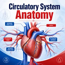


The heart is about the size of a closed fist, yet it beats roughly 100,000 times a day without any conscious effort from us. Its job is simple to state and remarkable to carry out: keep blood moving so that every tissue receives oxygen and nutrients and can get rid of waste. Understanding how the heart is built is one of the best starting points for anyone studying anatomy, nursing or other health-related fields.
Where the heart sits and what protects it
The heart lies in the chest, between the lungs, slightly tilted so that its pointed tip (the apex) angles toward the left side of the body. It is enclosed in a double-layered sac called the pericardium, which contains a small amount of lubricating fluid. This sac anchors the heart in place and lets it beat with very little friction.
The wall of the heart itself has three layers: the epicardium (the thin outer layer), the myocardium (the thick muscular middle layer that does the pumping) and the endocardium (a smooth lining that touches the blood). The myocardium is thicker on the left side because it must push blood to the entire body rather than just to the lungs.
The four chambers
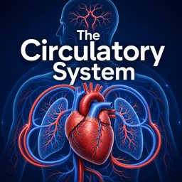
The heart is divided into four hollow chambers, two on the right and two on the left, separated by a muscular wall called the septum. Keeping the two sides separate means oxygen-poor blood never mixes with oxygen-rich blood.
- Right atrium: receives oxygen-poor blood returning from the body through two large veins, the superior and inferior vena cava.
- Right ventricle: pumps that blood to the lungs through the pulmonary artery.
- Left atrium: receives freshly oxygenated blood from the lungs through the pulmonary veins.
- Left ventricle: the strongest chamber, which pumps oxygen-rich blood into the aorta and out to the body.
The atria are the receiving chambers, while the ventricles are the main pumping chambers. This division of labor explains why the ventricle walls are much thicker than those of the atria.
The four valves
Valves act like one-way doors. They open to let blood pass forward and close to prevent it from flowing backward. The familiar “lub-dub” sound of a heartbeat comes largely from these valves closing.
| Valve | Location | Function |
|---|---|---|
| Tricuspid valve | Between the right atrium and right ventricle | Lets blood move into the right ventricle and stops backflow |
| Pulmonary valve | Between the right ventricle and the pulmonary artery | Directs blood to the lungs |
| Mitral valve | Between the left atrium and left ventricle | Lets oxygen-rich blood enter the left ventricle |
| Aortic valve | Between the left ventricle and the aorta | Sends blood out to the body |
Following the blood: two circuits
Blood travels through two connected loops. In the pulmonary circuit, oxygen-poor blood leaves the right ventricle, travels to the lungs, releases carbon dioxide and picks up oxygen, then returns to the left atrium. In the systemic circuit, the left ventricle sends this oxygen-rich blood through the aorta to the rest of the body. After delivering oxygen, it returns through the veins to the right atrium, and the cycle starts again.
Put in order, one trip looks like this:
- Body → vena cava → right atrium
- Right atrium → tricuspid valve → right ventricle
- Right ventricle → pulmonary valve → pulmonary artery → lungs
- Lungs → pulmonary veins → left atrium
- Left atrium → mitral valve → left ventricle
- Left ventricle → aortic valve → aorta → body
What makes the heart beat
The heart has its own built-in electrical system. A small cluster of specialized cells in the right atrium, called the sinoatrial (SA) node, acts as the natural pacemaker. It generates an electrical impulse that spreads across the atria, making them contract. The signal then reaches the atrioventricular (AV) node, which briefly delays it so that the ventricles have time to fill. After that, the impulse travels down special fibers through the septum and into the ventricle walls, triggering a coordinated contraction.
Each heartbeat has two main phases. During diastole, the heart relaxes and the chambers fill with blood. During systole, the ventricles contract and push blood out. The nervous system and hormones can speed up or slow down this rhythm depending on what the body needs, such as during exercise or rest.
The heart’s own blood supply
Although the heart is full of blood, its muscle cannot absorb nutrients directly from the chambers. Instead, it relies on the coronary arteries, which branch off the aorta and wrap around the heart’s surface. They deliver oxygen-rich blood to the myocardium. When these vessels become narrowed or blocked, the heart muscle can be deprived of oxygen, which is why coronary health is such an important topic in medicine and prevention.
Tips for studying heart anatomy
- Draw the heart repeatedly from memory, labeling chambers, valves and vessels.
- Color-code oxygen-rich blood (red) and oxygen-poor blood (blue) in your diagrams.
- Remember that “left” and “right” refer to the patient’s body, not to the viewer.
- Trace the blood path out loud, step by step, until it feels automatic.
- Connect structure to function: ask why each part has the shape and thickness it does.
Conclusion
The heart combines simple ideas into a highly efficient system: four chambers, four one-way valves, two circuits and an internal electrical conductor. Once you understand how these pieces fit together, topics such as blood pressure, heart sounds and common cardiovascular conditions become much easier to follow. If you want to keep building your knowledge, explore the related anatomy and health courses available on Cursa to continue learning at your own pace.