Velocity estimates are most reliable when the insonation angle is about 30° to 60°; errors increase substantially as the angle approaches 90°.

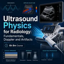
Duration of the online course: 6 hours and 8 minutes
New
Master ultrasound physics fast with a free online course: Doppler, artifacts, safety and image optimization for radiology and sonography—study anytime, earn skills.
Build a clear, practical understanding of ultrasound physics for radiology and everyday scanning decisions. This free online course bridges the gap between equations and what you see on the screen, so you can explain image behavior, choose better settings, and recognize why artifacts appear instead of guessing. Whether you are a student, technologist, resident, or a clinician who uses ultrasound at the point of care, you will gain the confidence to connect core physics to image quality, Doppler measurements, and safe scanning habits.
You will start with the fundamentals of sound waves and how frequency, wavelength, and propagation speed depend on the medium. From there, the course ties intensity and decibels to what matters clinically, including attenuation in soft tissue and why deeper structures demand different tradeoffs. You will see how pulse-echo imaging relies on timing and pulse parameters, and how reflection, refraction, scattering, and acoustic impedance shape what is transmitted back to the transducer. These principles quickly become intuitive tools for troubleshooting: understanding why interfaces look bright, why shadows form, and why angle and tissue type change the look of an exam.
The course also clarifies how transducers work and why design choices matter, from piezoelectric behavior to matching layers and damping. You will learn how common imaging modes create information differently, and how controls like time gain compensation help balance the image without distorting interpretation. As the course advances, you will connect beam geometry, focusing, steering, and spatial compounding to resolution tradeoffs, making it easier to justify choices that impact axial, lateral, elevational, and temporal resolution.
Doppler is covered in depth with a focus on real measurement quality: angle correction, preferred insonation angles, continuous versus pulsed wave Doppler, and what sets limits such as aliasing and the Nyquist relationship to PRF. You will also strengthen your ability to interpret spectral displays and understand how scale changes affect what you perceive. Finally, you will learn to identify frequent artifacts and the physics behind them, plus essential safety concepts including thermal and mechanical indices. Short quiz-style exercises reinforce key decisions, helping you translate physics into consistent, high-quality imaging practice.

6 hours and 8 minutes of online video course

Digital certificate of course completion (Free)

Exercises to train your knowledge

100% free, from content to certificate
How does the Doppler angle affect blood velocity measurements in ultrasound?
Velocity estimates are most reliable when the insonation angle is about 30° to 60°; errors increase substantially as the angle approaches 90°.
What causes aliasing in pulsed-wave Doppler ultrasound?
Aliasing occurs when the Doppler shift exceeds the Nyquist limit, which is one-half of the pulse repetition frequency (PRF).
What is the soft-tissue ultrasound attenuation rule of thumb?
In soft tissue, attenuation is approximately 0.5 dB per cm per MHz, increasing with both depth and frequency.
Ready to get started?Download the app and get started today.
Install the app now
to access the course
Over 5,000 free courses
Programming, English, Digital Marketing and much more! Learn whatever you want, for free.

Study plan with AI
Our app's Artificial Intelligence can create a study schedule for the course you choose.

From zero to professional success
Improve your resume with our free Certificate and then use our Artificial Intelligence to find your dream job.
You can also use the QR Code or the links below.

Free CourseOphthalmology
5

(7)

4h22m

15 exercises
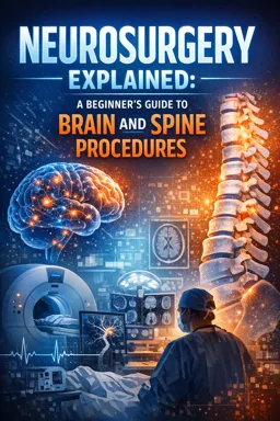
Free CourseNeurosurgery training
5
(2)
9h15m
13 exercises
Free CourseCadiology training
5
(5)
8h18m
14 exercises
Free CourseCongenital Heart Disease Echocardiography
5
(1)
21h23m
22 exercises

Free CourseMicrobiology
4.7

(11)
8h26m
7 exercises
Free CourseNeuro Pathology
4.7
(3)
16h55m
18 exercises
Free CourseRenal Pathology
4.7
(3)
14h06m
11 exercises
Free CourseECG Interpretation and Clinical Application: Electrocardiogram Essentials
3.8
(4)
9h21m
20 exercises
Free CourseEmbryology
New
12h03m
19 exercises
Carry knowledge in your pocket.
Download the Cursa app.
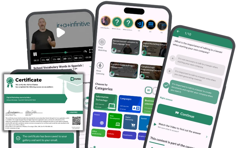
There are hundreds of free courses available, with a free certificate of completion that is saved in your mobile image gallery.
Download the app to access the Course Completion Certificate for Free.

+ 10 million
students

Free and Valid
Certificate

60 thousand free
exercises

4.8/5 rating in
app stores

Free courses in
video and ebooks