Duration of the online course: 21 hours and 23 minutes
New
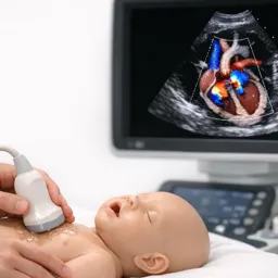
Build confidence reading congenital heart echoes with this free online course and quizzes—master key views, Doppler clues, and clinical decision support fast.
In this free course, learn about
Segmental analysis sequence for congenital heart disease (situs, AV, VA connections, associated lesions)
Normal transthoracic echo windows/views, including subxiphoid, parasternal, apical, and suprasternal imaging
Echo diagnosis of shunt lesions: ASD, VSD, AVSD, PDA, and aortopulmonary window (key views/clues)
Hypoplastic left heart syndrome: identifying restrictive atrial septal communication on echocardiography
Truncus arteriosus defining echo features and heterotaxy (asplenia/polysplenia) venous drainage patterns
Course Description
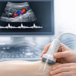
Congenital heart disease can look overwhelming on echocardiography, especially when anatomy is complex and small details change clinical decisions. This free online course is designed to help you develop a reliable, repeatable way to evaluate congenital lesions using transthoracic echo and Doppler, so your reports become clearer, your measurements more consistent, and your interpretations easier to defend in real cases.
You will start by strengthening the foundation: how to approach segmental analysis in the right sequence and how to recognize normal echocardiographic anatomy from standard windows. From there, the course guides you through the practical reasoning that separates a quick scan from a high-quality congenital study: choosing the best view for a specific question, identifying what each window can and cannot show, and using color and spectral Doppler to determine hemodynamic significance rather than relying on impressions.
Across a broad range of congenital diagnoses, you will practice the echo clues that matter at the bedside and in the reading room. You will sharpen your ability to assess shunts, valve and outflow tract abnormalities, arch pathology, venous connections, coronary anomalies, and complex conotruncal conditions, while paying attention to pitfalls such as misleading 2D measurements, flow aliasing, and orientation-dependent color findings. Clinical scenarios emphasize not just recognition, but what the finding implies for physiology, urgency, and next steps in imaging.
The learning experience is reinforced with questions and exercises that test interpretation and decision-making, helping you transform passive watching into active mastery. Whether you are a trainee building a framework, a sonographer refining technique, or a clinician who needs faster pattern recognition, this course supports a more confident approach to congenital echocardiography and clearer communication within the multidisciplinary team. Complete the activities to validate your progress and strengthen your day-to-day performance in congenital echo assessment.
Course content
Video class: Introduction53m
Exercise: In segmental analysis of congenital heart disease, what is the correct overall sequence of evaluation?
Video class: Chapter- 2 of 24 Normal echocardiographic views from different windows23m
Exercise: In the subxiphoid coronal (long-axis/anatomical) view, which pair of vessels is seen just anterior to the spine, with the inferior vena cava (IVC) closer to the transducer and the abdominal aorta farther away?
Video class: Chapter- 3 of 24 Echo evaluation of atrial septal defects1h43m
Exercise: In transthoracic echocardiography, which view is primarily used to visualize the superior (SVC) and inferior (IVC) rims of an ostium secundum atrial septal defect?
Video class: Chapter- 4 of 24 Echo evaluation of ventricular septal defects2h18m
Video class: Chapter- 5 of 24 Echo evaluation of atrioventricular septal defects55m
Exercise: Which feature best defines an intermediate atrioventricular septal defect (AVSD)?
Video class: Chapter- 6 of 24 Echo evaluation of patent ductus arteriosus56m
Exercise: Which echocardiographic view is primarily used to image the PDA from a high left parasternal window in a sagittal plane?
Video class: Chapter- 7 of 24 Echo evaluation of aortopulmonary window25m
Exercise: Which echocardiographic clue should raise suspicion for an aortopulmonary (AP) window as a cause of aortic runoff?
Video class: Chapter- 8 of 24 Left ventricular outflow tract anomalies52m
Exercise: Why can the aortic annulus measured on 2D echocardiography differ from the true anatomic aortic annulus?
Video class: Chapter- 9 of 24 Echo evaluation of aortic Coarctation28m
Exercise: In significant coarctation of the aorta, which Doppler feature is emphasized as a key sign of hemodynamically important obstruction?
Video class: Chapter- 10 of 24 Echo evaluation of Aortic arch anomalies and vascular rings32m
Exercise: In interrupted aortic arch, what structure must remain open to sustain blood flow to the descending aorta?
Video class: Chapter- 11 of 24 Right ventricular outflow tract obstruction33m
Exercise: How is valvular pulmonary stenosis severity classified by Doppler peak gradient?
Video class: Chapter- 12 of 24 Ebsteins anomaly of tricuspid valve37m
Exercise: In Epstein's anomaly, what echocardiographic finding indicates a significant apical displacement of the tricuspid valve leaflets?
Video class: Chapter- 13 of 24 Congenital Mitral valve anomalies34m
Exercise: Which congenital mitral valve lesion is defined by all mitral chordae attaching to a single papillary muscle, giving a parachute-like appearance and often an eccentric valve opening?
Video class: Chapter- 14 of 24 Coronary anomalies of clinical significance28m
Exercise: Which color Doppler finding is most characteristic of ALCAPA on echocardiography?
Video class: Chapter- 15 of 24 Echo evaluation of anomalies of Systemic veins41m
Exercise: Which echocardiographic finding suggests elevated right atrial pressure when assessing the inferior vena cava (IVC)?
Video class: Chapter- 16 of 24 Echo evaluation of anomalies of Pulmonary veins1h46m
Exercise: In a normal parasternal short-axis or suprasternal (crab) view, why do upper pulmonary veins typically appear blue and lower pulmonary veins red on color Doppler?
Video class: Chapter- 17 of 24 Tetralogy of Fallot53m
Exercise: What is the formula for the McGoon index used to assess pulmonary artery size in tetralogy of Fallot?
Video class: Chapter- 18 of 24 Double outlet right ventricle1h01m
Exercise: Which finding is part of the bilateral conus criterion for diagnosing double outlet right ventricle (DORV)?
Video class: Chapter- 19 of 24 D-Transposition of Great arteries1h35m
Exercise: What is the hallmark anatomical feature of transposition of the great arteries (TGA) on echocardiography?
Video class: Chapter- 20 of 24 L-Transposition of Great Arteries32m
Video class: Chapter- 21 of 24 Tricuspid atresia1h19m
Exercise: In tricuspid atresia type 1A (normally related great arteries), what is the primary source of pulmonary blood flow?
Video class: Chapter- 22 of 24 Hypoplastic left heart syndrome1h04m
Exercise: Which echocardiographic finding best indicates a restrictive interatrial communication in hypoplastic left heart syndrome?
Video class: Chapter- 23 of 24 Truncus arteriosus25m
Exercise: Which feature best defines truncus arteriosus on echocardiography?
Video class: Chapter-24 of 24 Heterotaxy syndromes - Asplenia and Polysplenia20m
Exercise: Which pulmonary venous drainage pattern is most typical in right isomerism (asplenia syndrome) compared with left isomerism (polysplenia syndrome)?