Duration of the online course: 4 hours and 22 minutes
5

(6)
Understanding eye complaints can be challenging because vision problems often sit at the intersection of medicine, neurology, and urgent care. This free online ophthalmology course is designed to help you connect those dots with clear, high-yield explanations and targeted practice questions. Whether you are studying for exams or strengthening clinical reasoning, you will develop a structured way to think through common presentations and the anatomy that makes them make sense.
You will start with essential concepts in glaucoma, focusing on what it is, why it damages the optic nerve, and how common drug classes work to reduce intraocular pressure by changing aqueous humor dynamics. From there, the course expands into cataracts with an emphasis on genetic associations, helping you recognize when an eye finding may be a clue to a broader syndrome.
A major strength of the course is its neuro-ophthalmology foundation. You will learn why the optic nerve is unique, how it fits into the central nervous system, and which cells create its myelin. You will also build a reliable mental model of the visual pathway, including where fibers cross and how lesions can translate into specific visual field patterns. The cranial nerves that move the eye are explained with practical clinical correlations, reinforcing how CN III, IV, and VI injuries present and why certain syndromes lead to characteristic deficits.
To keep your learning clinically relevant, the course links core mechanisms to real scenarios such as neonatal conjunctivitis patterns, trauma clues like periorbital ecchymosis, and microvascular complications related to diabetes. It also highlights time-sensitive conditions where fast recognition matters, including giant cell arteritis and its diagnostic cues that warrant immediate action. By the end, you will be better prepared to interpret symptoms, localize problems, and communicate eye findings with confidence in study settings or patient care.
4 hours and 22 minutes of online video course
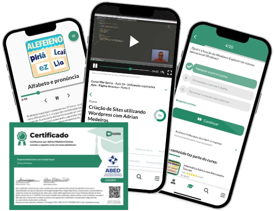
Digital certificate of course completion (Free)
Exercises to train your knowledge
100% free, from content to certificate
Ready to get started?Download the app and get started today.
Install the app now
to access the courseOver 5,000 free courses
Programming, English, Digital Marketing and much more! Learn whatever you want, for free.
Study plan with AI
Our app's Artificial Intelligence can create a study schedule for the course you choose.
From zero to professional success
Improve your resume with our free Certificate and then use our Artificial Intelligence to find your dream job.
You can also use the QR Code or the links below.


Free CourseNeurosurgery training
5
(1)

9h15m

13 exercises

Free CourseCadiology training
5
(5)
8h18m
14 exercises
Free CourseUltrasound Physics for Radiology: Fundamentals, Doppler and Artifacts
5
(1)
6h08m
24 exercises

Free CourseMicrobiology
4.78
(9)
8h26m
7 exercises

Free CourseNeuro Pathology
4.67

(3)
16h55m
18 exercises

Free CourseRenal Pathology
4.5
(2)
14h06m
11 exercises
Free CourseECG Interpretation and Clinical Application: Electrocardiogram Essentials
1
(1)
9h21m
20 exercises

Free CourseEmbryology
New
12h03m
19 exercises
Free CourseCongenital Heart Disease Echocardiography
New
21h23m
22 exercises
Thousands of online courses in video, ebooks and audiobooks.
To test your knowledge during online courses
Generated directly from your cell phone's photo gallery and sent to your email
Download our app via QR Code or the links below::.
Carry knowledge in your pocket.
Download the Cursa app.
There are hundreds of free courses available, with a free certificate of completion that is saved in your mobile image gallery.
Download the app to access the Course Completion Certificate for Free.

+ 10 million
students
Free and Valid
Certificate
60 thousand free
exercises
4.8/5 rating in
app stores
Free courses in
video and ebooks