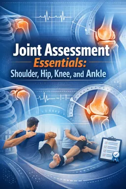
Why add strength testing after ROM?
Range of motion (ROM) tells you how far a joint moves and what limits it. Basic strength testing tells you how well the tissues can produce and tolerate force in that position. When paired, ROM + strength helps you distinguish “can’t move” from “can’t control,” and “stiff” from “irritable.”
This chapter focuses on beginner-friendly strength testing that complements ROM findings: manual muscle testing (MMT) and simple resisted isometrics. The goal is not to diagnose a specific pathology from one test, but to document a repeatable baseline and identify patterns (painful vs painless weakness; side-to-side differences; which movement directions are most limited).
MMT and resisted isometrics: purpose and limits
What these tests are good for
- Screening contractile function: can the muscle-tendon unit generate force in a specific direction?
- Comparing sides: right vs left under similar positioning and resistance.
- Tracking change: before/after an intervention or over time.
- Clarifying symptoms: does pain appear when the patient contracts against resistance?
Key limits to remember
- They are not “pure muscle tests.” Pain, fear, coordination, and joint irritation can reduce force output.
- Hand-held resistance is variable. Your strength and leverage affect results; prioritize consistency over “max force.”
- MMT is position-specific. A muscle can test “strong” in one position and “weak” in another due to length, pain, or stabilization differences.
- Isometrics don’t test dynamic control. A strong isometric does not guarantee good movement quality during functional tasks.
Grading in plain language (with pain notes)
If you do not use formal 0–5 grades, use a simple, repeatable scale and always record pain separately.
| Label | What it looks like | How to document |
|---|---|---|
| Strong | Holds position against your maximal safe resistance without giving way | Strong, no pain or Strong, pain 4/10 |
| Moderate | Holds against some resistance but gives way with higher resistance | Moderate, pain at end of hold |
| Weak | Cannot hold against even light resistance or cannot reach test position | Weak, no pain or Weak, pain-limited |
Pain recording rule: document location and intensity (e.g., 0–10) and whether pain appears immediately or after a few seconds. Example: Hip abd: Moderate, lateral hip pain 5/10 within 2s.
Core technique: how to perform a clean resisted isometric/MMT
Step-by-step sequence
- Choose a test position that is comfortable, stable, and matches your goal (mid-range for general strength; specific angles if symptoms are angle-dependent).
- Explain the task in one sentence: “Hold this position; don’t let me move you.”
- Set the limb in the desired direction and ensure the patient is not already compensating (trunk lean, shoulder hike, pelvic rotation).
- Stabilize proximally (scapula, pelvis, thigh) so the force comes from the target region rather than from substitution.
- Apply resistance gradually over ~1–2 seconds to a firm “test” level, then hold ~3–5 seconds.
- Watch for the first failure: giving way, substitution, breath-holding, facial grimace, or pain report.
- Ask two quick questions: “Where did you feel it?” and “Was it pain or effort?”
- Repeat once if the first attempt looked uncoordinated. Keep total repetitions low if irritable.
Choosing positions: practical rules
- Mid-range first when the goal is baseline strength and symptom irritability is unknown.
- Short-lever first (resistance closer to the joint) if pain is high or control is poor; progress to long-lever for more challenge.
- Reduce gravity demand (side-lying, supine) if the patient cannot even get into position against gravity.
- Match the complaint: if pain occurs in a specific ROM region, test isometrics near that angle (within tolerance) to see if symptoms are contractile.
Stabilization and resistance placement
Two common reasons for “false weakness” are poor stabilization and resistance applied too far from the joint too soon.
- Listen to the audio with the screen off.
- Earn a certificate upon completion.
- Over 5000 courses for you to explore!

Download the app
- Stabilize the segment above the joint: scapula for shoulder tests; pelvis for hip tests; femur for knee tests; tibia for ankle tests.
- Place resistance distal to the joint for a stronger test, but move it more proximal if pain or leverage overwhelms the patient.
- Keep your line of force opposite the movement (e.g., for shoulder external rotation, resist into internal rotation).
Recording: separate pain from weakness
Use a consistent template so your notes are comparable across visits.
Movement: position / lever / stabilization used / result / pain details / substitutionsExample:
Shoulder ER: sitting, elbow 90 at side; stabilize humerus; resist at distal forearm; Moderate; anterior shoulder pain 3/10 at 4s; mild trunk rotationJoint-specific test menus (beginner-friendly)
Use these as “menus,” not mandatory checklists. Pick the tests that best match the ROM findings and the patient’s symptoms.
Shoulder: rotator cuff and deltoid patterns
1) External rotation (infraspinatus/teres minor bias)
- Position: Sitting or standing, elbow flexed 90°, arm at side, towel between elbow and trunk if needed.
- Stabilize: Keep elbow tucked; stabilize distal humerus to prevent abduction.
- Action: “Rotate your forearm outward; hold.”
- Resistance: At distal forearm into internal rotation.
- Watch for: Trunk rotation, shoulder abduction, wrist extension.
2) Internal rotation (subscapularis bias)
- Position: Sitting/standing, elbow 90°, arm at side.
- Stabilize: Prevent shoulder extension and trunk rotation.
- Action: “Rotate your forearm inward; hold.”
- Resistance: At distal forearm into external rotation.
- Note: If anterior shoulder pain is provoked, record whether pain is deep/anterior and whether strength remains strong.
3) Abduction (deltoid/supraspinatus pattern)
- Beginner option (safer leverage): Test at ~30–60° abduction in the scapular plane (slightly forward of pure side abduction).
- Position: Sitting or standing; arm slightly forward of the body.
- Stabilize: Prevent shoulder hiking; monitor scapular elevation.
- Action: “Lift out and slightly forward; hold.”
- Resistance: At distal humerus (short lever) or near elbow; progress toward wrist only if tolerated.
- Watch for: Upper trapezius dominance, trunk lean.
4) Flexion (anterior deltoid pattern)
- Position: Sitting/standing, shoulder flexed ~60–90° (as tolerated), elbow slightly bent.
- Stabilize: Prevent trunk extension/lean back.
- Resistance: Downward at distal humerus/forearm.
5) Extension (posterior deltoid/lat pattern)
- Position: Prone with arm by side or standing with shoulder slightly extended.
- Stabilize: Prevent trunk rotation and scapular retraction substitution.
- Resistance: Toward flexion at distal humerus.
Hip: abductors and extensors
1) Hip abduction (gluteus medius/minimus pattern)
- Position (preferred): Side-lying, test leg on top, hip neutral or slight extension, knee straight.
- Stabilize: Pelvis—prevent rolling backward; keep trunk stacked.
- Action: “Lift your leg toward the ceiling; hold. Don’t roll your hips.”
- Resistance: Downward at distal femur (above knee) first; progress toward ankle if appropriate.
- Watch for: Hip flexion (TFL substitution), pelvic rotation, hiking.
2) Hip extension (gluteus maximus pattern)
- Position (beginner-friendly): Prone with knee bent to ~90° to reduce hamstring dominance, hip in neutral.
- Stabilize: Pelvis—prevent lumbar extension/anterior pelvic tilt.
- Action: “Lift your thigh slightly off the table; hold.”
- Resistance: Downward at distal femur.
- Watch for: Low back extension, pelvic rotation.
Alternative if prone is not tolerated: Standing hip extension with hands supported on a table; resist at posterior thigh while preventing trunk lean.
Knee: extensors and flexors
1) Knee extension (quadriceps pattern)
- Position: Sitting at edge of table/chair, knee flexed ~90°.
- Stabilize: Femur—avoid hip hiking; ensure patient is not leaning back to assist.
- Action: “Straighten your knee; hold.”
- Resistance: At distal tibia (above ankle) into knee flexion.
- Watch for: Hip flexion/lean back, ankle plantarflexion bracing.
2) Knee flexion (hamstrings pattern)
- Position: Prone with knee flexed ~45–70° (or sitting if prone not tolerated).
- Stabilize: Pelvis—prevent hip flexion/rotation.
- Action: “Bend your knee; hold.”
- Resistance: At distal tibia into knee extension.
- Watch for: Hip extension substitution, cramping.
Ankle: dorsiflexors, plantarflexors, invertors, evertors
General setup tips for ankle tests
- Position: Sitting with lower leg supported and foot off edge, or long-sitting.
- Stabilize: Distal tibia/fibula to reduce knee/hip substitution.
- Foot position: Start near neutral; avoid testing from extreme plantarflexion/inversion if irritable.
1) Dorsiflexion (tibialis anterior pattern)
- Action: “Pull your foot up toward you; hold.”
- Resistance: Over dorsum/medial forefoot into plantarflexion.
- Watch for: Toe extension substitution (EHL), foot inversion.
2) Plantarflexion (gastroc/soleus pattern)
- Seated isometric option: Ankle neutral; “Press down like a gas pedal; hold.” Resist at plantar forefoot into dorsiflexion.
- Functional option (if appropriate): Single-leg heel raise count/quality for a quick endurance screen; document pain and height symmetry.
- Watch for: Midfoot collapse, knee flexion strategy, reduced heel height.
3) Inversion (tibialis posterior/anterior pattern)
- Action: “Turn the sole inward; hold.”
- Resistance: At medial forefoot into eversion.
- Watch for: Toe curling, tibial rotation.
4) Eversion (peroneals pattern)
- Action: “Turn the sole outward; hold.”
- Resistance: At lateral forefoot into inversion.
- Watch for: Hip external rotation substitution, toe extension.
Interpretation rules you can apply immediately
1) Pain with strong force
Pattern: The patient can hold strong resistance, but it reproduces pain.
Most consistent with: contractile irritation (muscle-tendon unit is sensitive but still capable of producing force).
How to document: Strong, pain 5/10 plus location and timing.
2) Weakness without pain
Pattern: Clear giving way or inability to hold, but no pain is reported.
Most consistent with: inhibition, deconditioning, or motor control deficit (also consider neurological contribution if weakness is marked or widespread).
Next practical step: Re-check stabilization and lever length; if still weak, compare to functional tasks and consider additional screening as appropriate.
3) Weakness with pain
Pattern: The patient reports pain and cannot sustain resistance.
Most consistent with: combined impairment (pain-limited force output and reduced capacity).
Documentation tip: Specify whether weakness appears to be pain-limited (stops because it hurts) versus true giving way (cannot hold even when pain is minimal).
Quick cross-checks to improve confidence
- Change lever arm: If long-lever is weak/painful, retest short-lever. A big improvement suggests leverage/pain sensitivity rather than global failure.
- Change position: If a test is painful in one position, try a supported position (e.g., side-lying hip abduction vs standing). Consistent pain across positions supports contractile sensitivity.
- Compare directions: A single-direction deficit (e.g., shoulder ER weaker than IR) is more informative than “everything weak.”