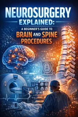
In practical terms, neurosurgery is the specialty that evaluates and treats conditions affecting the brain, spinal cord, spine (the bones and discs that protect the cord and nerve roots), and peripheral nerves (nerves outside the brain and spinal cord). A neurosurgeon’s work includes deciding when an operation is helpful, performing procedures when needed, and coordinating non-surgical care when surgery is not the best option.
What neurosurgeons treat (and how that differs from related specialties)
Neurosurgery: typical scope
- Brain: tumors, bleeding (hemorrhage), hydrocephalus/CSF flow problems, traumatic injuries, vascular problems (e.g., aneurysms, AVMs), some epilepsy surgery, functional procedures (e.g., deep brain stimulation in selected cases).
- Spine and spinal cord: disc herniations, spinal stenosis, fractures/instability, spinal tumors, infections, spinal cord compression.
- Peripheral nerves: nerve entrapments (e.g., carpal tunnel), nerve tumors, traumatic nerve injuries, selected pain-related nerve procedures.
How roles commonly differ in real clinics
| Specialty | Common focus | Typical “handoff” to neurosurgery |
|---|---|---|
| Neurology | Diagnosis and medical management of nervous system disorders (e.g., migraine, epilepsy meds, MS, neuropathy) | Structural lesion on imaging, progressive neurological deficit, refractory seizures needing surgical evaluation |
| Orthopedics (spine) | Musculoskeletal spine problems; many spine surgeries overlap with neurosurgery | Complex spinal cord/nerve compression, intradural tumors, certain deformity/instability cases depending on local practice |
| Pain management | Non-surgical pain control (medications, injections, nerve blocks, neuromodulation trials) | Clear compressive cause needing decompression, or implantable neuromodulation in collaboration (e.g., spinal cord stimulation) |
| Rehabilitation (PM&R), PT/OT | Function, recovery, mobility, strengthening, adaptive strategies | Worsening weakness, new bowel/bladder issues, or symptoms not improving as expected prompting re-evaluation |
In many systems, the key practical distinction is: neurosurgeons are asked to judge whether a structural problem is present and whether an intervention on the brain/spine/nerves will improve safety, function, or quality of life.
(1) Common symptom pathways: what symptoms can indicate
Symptoms are “pathways” rather than diagnoses. The same symptom can come from benign causes or from conditions that threaten the brain or spinal cord. The goal is to connect the symptom pattern to the most likely anatomical location and urgency.
Headache
- Often non-surgical: migraine, tension-type headache, medication overuse, sinus-related pain.
- Can indicate neurosurgical disease when paired with red flags: sudden worst headache, headache with neurological deficits, headache with fever/neck stiffness, new headache in someone with cancer/immunosuppression, headache with progressive morning vomiting or worsening when lying down (possible raised intracranial pressure).
Practical pathway: headache pattern + neurological exam → decide if urgent imaging is needed (CT/MRI) → if imaging shows bleeding, mass effect, hydrocephalus, or infection-related collections, neurosurgery becomes involved.
Weakness (arm/leg/face) or clumsiness
- Brain causes: stroke/bleed, tumor, abscess, subdural hematoma.
- Spinal cord causes: cervical/thoracic cord compression (stenosis, tumor, epidural abscess/hematoma).
- Nerve root/peripheral nerve causes: radiculopathy (e.g., L5 weakness), plexopathy, entrapment.
Practical pathway: determine distribution (one side vs both legs, proximal vs distal) → check for upper motor neuron signs (spasticity, hyperreflexia) suggesting cord/brain → urgent imaging if acute or progressive → surgery considered if there is compressive pathology with deficit.
- Listen to the audio with the screen off.
- Earn a certificate upon completion.
- Over 5000 courses for you to explore!

Download the app
Numbness/tingling
- Peripheral patterns: hand numbness in median nerve distribution (carpal tunnel), stocking-glove neuropathy (often medical).
- Radicular patterns: dermatomal numbness with neck/back pain radiating to arm/leg.
- Spinal cord patterns: numbness in both hands, band-like trunk sensation, sensory level on the torso.
Practical pathway: map the sensory complaint (dermatome vs nerve vs “level”) → correlate with exam → imaging and/or nerve testing as appropriate → neurosurgical input when imaging shows nerve root or cord compression that matches symptoms.
Back pain and neck pain
- Common non-surgical: muscle strain, degenerative disc disease without nerve compression, facet-related pain.
- Potentially neurosurgical when: pain radiates with weakness/numbness (radiculopathy), progressive walking difficulty (myelopathy), pain after trauma with instability, pain with fever or IV drug use (possible infection), pain with cancer history (possible metastasis).
Practical pathway (step-by-step):
- Step 1: Screen for red flags (trauma, fever, cancer, new weakness, bowel/bladder changes).
- Step 2: Identify nerve involvement (radiating pain below knee/into arm, dermatomal numbness, reflex changes).
- Step 3: Start conservative care when appropriate (activity modification, PT, anti-inflammatory strategy if safe, targeted injections when indicated).
- Step 4: Reassess at a defined interval (often weeks) or sooner if worsening.
- Step 5: Obtain MRI/CT when deficits, red flags, or failure of conservative therapy occur.
- Step 6: Consider surgery if imaging shows a surgically correctable cause that matches symptoms and goals.
Seizures
- Often managed medically first: anti-seizure medications and neurologic evaluation.
- Neurosurgical involvement when: imaging shows a lesion (tumor, vascular malformation, scar), seizures are refractory to medications, or there is a need for biopsy/resection or invasive monitoring.
Practical pathway: confirm seizure type and triggers → MRI/EEG → treat medically → if seizures persist or a structural cause is found, neurosurgery evaluates whether removing/altering the lesion or performing epilepsy surgery could reduce seizures.
Gait imbalance, frequent falls, or “heavy legs”
- Brain causes: normal pressure hydrocephalus pattern (gait-first), posterior fossa lesions, stroke.
- Spinal cord causes: cervical myelopathy (often with hand clumsiness), thoracic cord compression.
- Non-surgical contributors: inner ear disorders, medication effects, neuropathy, deconditioning.
Practical pathway: characterize gait (wide-based, shuffling, spastic) → look for cord signs (hyperreflexia, Hoffmann sign, Babinski) → MRI brain and/or spine based on exam → neurosurgical decisions focus on whether there is progressive cord/brain compression where delay risks permanent deficit.
(2) Decision framework: surgery vs non-surgical care
Neurosurgical decision-making is less about “can we operate?” and more about “will an operation improve outcomes compared with non-surgical care, and what is the risk of waiting?” A practical framework uses five recurring questions.
Question 1: Is there a threat to life, brain, spinal cord, or nerve function?
- Examples: expanding brain bleed, ruptured aneurysm, acute hydrocephalus, spinal epidural abscess with deficits, cauda equina syndrome.
- Implication: surgery or urgent intervention is favored because delay can cause irreversible injury or death.
Question 2: Are there objective neurological deficits?
- Deficits that raise urgency: progressive weakness, new speech difficulty, worsening gait from myelopathy, bowel/bladder dysfunction, loss of hand function.
- Why it matters: deficits suggest the nervous system is already compromised; earlier decompression or treatment may improve recovery potential.
Question 3: Do imaging findings match the symptoms and exam?
Imaging abnormalities are common, especially in the spine. Surgery is most effective when there is a clear clinic–imaging match.
- Good match example: right L5 weakness + right L4-5 disc herniation compressing the L5 nerve root.
- Poor match example: diffuse back pain with mild degenerative changes but no nerve compression and a normal neurological exam.
Question 4: Has appropriate conservative therapy been tried (when safe)?
Many conditions improve without surgery. “Conservative therapy” is not doing nothing; it is a structured plan with a time horizon.
- Typical components: physical therapy, activity modification, anti-inflammatory strategy if appropriate, neuropathic pain medications when indicated, targeted injections, lifestyle and ergonomic changes.
- Failure definition (practical): persistent disabling symptoms after an adequate trial, inability to return to essential activities, or worsening deficits despite treatment.
Question 5: Do the expected benefits outweigh the risks for this patient?
This is individualized. Two people with the same MRI can make different reasonable choices.
- Benefit types: prevent deterioration (e.g., cord compression), restore function (e.g., relieve nerve root compression), reduce seizure burden, obtain diagnosis (biopsy), relieve pressure (hydrocephalus).
- Risk types: anesthesia risk, infection, bleeding, CSF leak, neurological worsening, need for reoperation, adjacent-segment degeneration (spine), incomplete symptom relief.
Step-by-step decision workflow used in many neurosurgical visits:
- Step 1: Define the main problem in one sentence (e.g., “progressive hand clumsiness from cervical cord compression”).
- Step 2: Confirm objective findings (exam deficits, reflex changes, gait testing).
- Step 3: Review imaging together and identify the target (what would be decompressed/removed/stabilized).
- Step 4: List reasonable options: observation, medical/rehab, injections, surgery (and which type).
- Step 5: Compare outcomes: what improves, what may not improve, and what happens if you wait.
- Step 6: Align with patient goals and constraints (work demands, caregiving, tolerance for risk, recovery time).
- Step 7: Decide timing and next steps (pre-op optimization, second opinion, or conservative plan with follow-up triggers).
(3) What “emergent,” “urgent,” and “elective” mean in neurosurgery
These terms describe time sensitivity based on risk of irreversible harm, not how “serious” something feels.
Emergent
Meaning: needs immediate action (often hours) because delay risks death or permanent neurological injury.
- Examples: large intracranial hemorrhage with mass effect, acute obstructive hydrocephalus, traumatic epidural hematoma, spinal cord compression with rapidly worsening deficits, cauda equina syndrome with urinary retention.
- Typical pathway: emergency department → rapid imaging → neurosurgical decision → OR/intervention or ICU-level monitoring.
Urgent
Meaning: should be addressed soon (days to a couple of weeks) because function is at risk, symptoms are progressing, or the window for best recovery may close.
- Examples: progressive cervical myelopathy, brain tumor with worsening deficits but stable vital status, spinal infection requiring decompression/drainage, symptomatic subdural hematoma without rapid decline.
- Typical pathway: expedited clinic/inpatient evaluation → pre-op workup → scheduled surgery soon, or close monitoring with clear escalation criteria.
Elective
Meaning: planned in advance (weeks to months) because it is safe to schedule and optimize the patient, and waiting is unlikely to cause permanent harm in the short term.
- Examples: stable lumbar stenosis causing activity-limiting leg pain, certain benign tumors without deficits, carpal tunnel release after conservative measures, selected spine stabilization for chronic instability.
- Typical pathway: outpatient evaluation → trial of conservative care when appropriate → shared decision-making → scheduled surgery with prehabilitation/optimization.
Practical tip: “Elective” does not mean “optional” in the everyday sense; it means “not time-critical today.” Some elective surgeries are still strongly recommended to prevent gradual decline.
(4) Multidisciplinary care and shared decision-making
Neurosurgical care often involves a team because nervous system problems can affect movement, speech, cognition, pain, and daily function. Teams also reduce blind spots: one specialist may see a surgical target, another may see a better non-surgical route.
Who may be involved (and what they contribute)
- Neurosurgeon: determines surgical candidacy, explains procedure options, risks, and expected outcomes; coordinates perioperative plan.
- Neurologist: diagnostic refinement (e.g., seizure type, neuropathy vs radiculopathy), medical management, long-term follow-up for non-surgical conditions.
- Radiologist: interprets imaging; in complex cases, neuroradiology input can clarify subtle findings.
- Anesthesiology and perioperative medicine: risk assessment, optimization of comorbidities, pain control strategy.
- Oncology/radiation oncology: for tumors—systemic therapy, radiation planning, sequencing with surgery.
- Infectious disease: for abscess/osteomyelitis—antibiotic selection and duration.
- Rehabilitation (PT/OT/SLP): functional baseline, recovery planning, gait and balance training, adaptive equipment.
- Pain management: injections, medication strategies, neuromodulation evaluation, chronic pain support.
How shared decision-making works in practice
Shared decision-making means the clinician contributes medical expertise and the patient contributes values, goals, and acceptable trade-offs. A useful way to structure the conversation is to make goals explicit and connect them to measurable outcomes.
Step-by-step shared decision checklist:
- Step 1: Clarify the patient’s top goals (examples: “walk 20 minutes,” “return to work,” “avoid opioids,” “reduce seizure frequency,” “preserve hand function”).
- Step 2: Translate goals into clinical targets (pain relief vs strength recovery vs preventing decline).
- Step 3: Review options side-by-side (including doing nothing) with expected probabilities when known.
- Step 4: Discuss what surgery can and cannot fix (e.g., decompression may relieve leg pain faster than it reverses long-standing numbness).
- Step 5: Identify personal risk factors (smoking, diabetes control, bone density, anticoagulants, frailty) and whether optimization can reduce risk.
- Step 6: Decide on timing and “tripwires” for escalation if choosing non-surgical care (e.g., new weakness, falls, bladder changes, worsening gait).
Quality-of-life considerations that often drive decisions
- Function over imaging: the decision often hinges on what the patient cannot do, not how dramatic the MRI looks.
- Recovery time and support: home help, job demands, transportation, and caregiving responsibilities can influence timing and procedure choice.
- Risk tolerance: some patients prioritize avoiding neurological decline; others prioritize avoiding surgical risk unless clearly necessary.
- Chronic symptoms: longer-standing nerve damage may improve incompletely; setting expectations prevents disappointment and helps choose the right option.
Practical example (spine): Two patients have similar lumbar stenosis. Patient A’s goal is to walk without stopping and has failed PT and injections; surgery may offer meaningful functional improvement. Patient B has mild symptoms controlled with activity modification and prefers to avoid downtime; continued conservative care with clear follow-up triggers may be best.
Practical example (brain lesion): A small incidental meningioma without symptoms may be monitored with serial imaging. A lesion causing seizures or progressive weakness shifts the balance toward intervention because the symptom burden and neurological risk are higher.