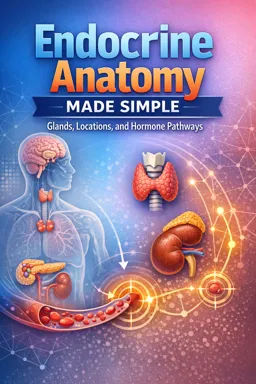
How to Use an Integrated Endocrine Map
This chapter merges all major endocrine gland locations into one repeatable “map-and-pathway” template so you can predict where a hormone enters the circulation and how it distributes to the rest of the body. You will build a cumulative diagram by body region using the same four-part scaffold each time:
- Gland position (where it sits)
- Nearby landmark (what you can reliably find next to it)
- Primary venous outflow direction (the first venous route it uses)
- Major targets (high-yield destinations after systemic distribution)
Use the same visual template repeatedly. On paper (or a tablet), draw a simple body outline divided into four regions: cranial, cervical, abdominal, pelvic. For each gland, add a small icon, then draw a short arrow to the nearest landmark, then a thicker arrow showing the first venous outflow, then thin branching arrows to major targets.
Reusable Visual Template (Copy/Paste for Each Gland)
[Gland icon] → (Nearest landmark) → {First venous outflow} ⇒ [Major targets]Keep the arrows consistent: → for local relationships, {} for the first venous route, and ⇒ for whole-body distribution.
Cranial Region: Hypothalamus–Pituitary as the “Headwater” Node
In your integrated map, treat the cranial endocrine structures as the upstream control node that influences multiple downstream glands. You are not re-learning detailed pituitary microanatomy here; you are placing it into the whole-body distribution logic.
| Template item | What to draw/label |
|---|---|
| Gland position | Midline cranial base (sellar region) |
| Nearby landmark | Sphenoid/sellar region; optic chiasm region (as a spatial cue) |
| Primary venous outflow direction | Venous drainage into dural venous sinuses (cranial venous system) |
| Major targets | Thyroid, adrenal cortex, gonads, breast, kidney (via downstream endocrine axes) |
Diagram step: Place a small “control node” icon in the cranial region. Draw a short arrow to a skull-base landmark, then a thick arrow to “cranial venous sinuses,” then branching arrows to the cervical (thyroid), abdominal (adrenals/pancreas), and pelvic (gonads) regions to emphasize axis-level influence.
- Listen to the audio with the screen off.
- Earn a certificate upon completion.
- Over 5000 courses for you to explore!

Download the app
Guided Case Prompts (Cranial)
- Prompt 1: A hormone released from the sellar region enters which venous compartment first: a neck vein, a portal vein, or a dural venous sinus system?
- Prompt 2: If a pituitary hormone targets the adrenal cortex, which body region will you trace to next on your map?
- Prompt 3: When you draw “major targets,” do you draw one target organ or multiple? (Answer by pointing to at least three regions.)
Cervical Region: Thyroid–Parathyroid Cluster as a Neck Distribution Hub
On the integrated map, the cervical region is your “neck hub” where endocrine output rapidly joins systemic venous return. You already know the gland-specific landmarks; here you will connect them to the first venous outflow and then to major targets.
| Gland | Gland position → landmark | Primary venous outflow direction | Major targets (high-yield) |
|---|---|---|---|
| Thyroid | Anterior neck around upper trachea → larynx/trachea as landmark | Thyroid veins → internal jugular/brachiocephalic venous pathways (systemic) | Most tissues (metabolic rate), heart, brain, skeletal muscle |
| Parathyroids | Posterior thyroid region → posterior thyroid surface as landmark | Venous drainage follows thyroid venous routes (systemic) | Bone, kidney, indirectly gut (via calcium/phosphate regulation) |
Diagram step (cumulative): In the cervical region of your body outline, draw the thyroid “butterfly” icon and small dots for parathyroids. Add a short arrow to the trachea/larynx landmark. Then draw a thick arrow labeled “thyroid veins → central venous return.” Finally, branch thin arrows to “heart,” “skeletal muscle,” “brain,” and “bone/kidney.”
Guided Case Prompts (Cervical)
- Prompt 1: A hormone released from the thyroid enters which route first: a lymphatic duct, a thyroid vein, or the portal vein?
- Prompt 2: If venous outflow from the neck is impaired, which direction on your map would you expect congestion to affect first: cranial, thoracic/central venous, or pelvic?
- Prompt 3: Parathyroid hormone reaches bone and kidney quickly. On your diagram, do you route it through a special portal system or through standard systemic venous return?
Abdominal Region: Retroperitoneal and Portal-Adjacent Endocrine Sources
The abdominal region is where learners often lose the “first venous step” because multiple drainage patterns exist (systemic caval routes vs portal routes). Your integrated map should make the first venous outflow explicit for each gland.
Adrenal (Suprarenal) Glands: Systemic Venous Outflow to Central Circulation
| Template item | What to draw/label |
|---|---|
| Gland position | Superior to kidneys (retroperitoneal) |
| Nearby landmark | Upper pole of each kidney; diaphragm/upper retroperitoneum |
| Primary venous outflow direction | Adrenal veins → renal vein/IVC pathway (systemic; side-specific details can be added if you already know them) |
| Major targets | Vascular smooth muscle, liver, immune tissues, many organs (stress and electrolyte-related targets) |
Diagram step: Place two small triangles above the kidneys. Draw a short arrow to the kidney upper pole. Then draw a thick arrow to “renal vein/IVC → heart.” Then branch to “vessels,” “liver,” “immune tissues,” and “kidney.”
Pancreatic Islets: Portal-First Logic for Liver-First Exposure
In an integrated map, the pancreas is the key example of a gland whose hormones often have a prominent early effect on the liver because venous drainage from much of the gastrointestinal region flows to the portal system before returning to the heart.
| Template item | What to draw/label |
|---|---|
| Gland position | Upper abdomen, posterior to stomach (retroperitoneal/secondarily retroperitoneal portions) |
| Nearby landmark | Stomach/duodenum region; splenic hilum direction for tail |
| Primary venous outflow direction | Pancreatic venous drainage → portal venous system → liver (portal-first) |
| Major targets | Liver (early/high exposure), skeletal muscle, adipose tissue |
Diagram step: Draw an elongated pancreas shape. Add a short arrow to “posterior to stomach/near duodenum.” Then draw a thick arrow to “portal vein → liver.” From the liver, draw a second thick arrow to “hepatic veins → IVC → heart” to show the two-step venous sequence. Then branch thin arrows to “muscle” and “adipose.”
Guided Case Prompts (Abdominal)
- Prompt 1: A hormone released from pancreatic islets will enter which venous route first on your diagram: portal venous system or IVC?
- Prompt 2: A hormone released from an adrenal gland reaches the heart by which first labeled venous route on your map?
- Prompt 3: If the liver is a major early target, which gland’s pathway should visibly pass through the portal system before reaching systemic circulation?
Pelvic Region: Gonadal Endocrine Output and Systemic Distribution
In the pelvic region, your integrated map should emphasize that gonadal hormones enter venous drainage and then distribute widely, with major effects on reproductive organs and many non-reproductive tissues (bone, muscle, brain).
| Gland | Gland position → landmark | Primary venous outflow direction | Major targets (high-yield) |
|---|---|---|---|
| Ovaries | Lateral pelvic cavity → uterus/uterine tubes as landmark | Ovarian veins → systemic venous return (to central venous circulation) | Uterus/breast, bone, brain, cardiovascular system |
| Testes | Scrotum → spermatic cord/inguinal canal as landmark | Pampiniform plexus → testicular veins → systemic venous return | Reproductive tract, muscle, bone, brain |
Diagram step: In the pelvic region, place paired gonad icons (ovaries in pelvis; testes in scrotum). Draw a short arrow to the nearest landmark (uterus/tubes for ovaries; spermatic cord for testes). Then draw a thick arrow to “gonadal veins → central venous return.” Finally, branch thin arrows to “bone,” “muscle,” “brain,” and “reproductive organs.”
Guided Case Prompts (Pelvic)
- Prompt 1: A hormone released from the testes enters which structure first on your pathway drawing: pampiniform plexus/testicular veins or portal vein?
- Prompt 2: When tracing ovarian hormone distribution, which major non-reproductive target should you include to show whole-body effects?
- Prompt 3: If you had to choose one “direction” arrow for gonadal hormones after venous outflow, would it be toward the liver first (portal) or toward central venous return (systemic)?
Putting It All Together: One Cumulative Whole-Body Diagram
Now combine all regions into a single integrated map. The goal is not artistic detail; it is consistent placement and correct first-venous-step logic.
Step-by-Step Build (Cumulative Diagram)
- Step 1 (Frame): Draw a simple body outline and divide it into four labeled zones: cranial, cervical, abdominal, pelvic.
- Step 2 (Place glands): Add icons in each zone: cranial control node; thyroid/parathyroids; adrenals and pancreas; gonads.
- Step 3 (Add landmarks): Next to each icon, add one nearby landmark label (sellar/skull base; trachea/larynx; kidney upper pole and stomach/duodenum; uterus/tubes or spermatic cord).
- Step 4 (First venous outflow): Draw one thick arrow from each gland to its first venous route label. Use only these route labels to keep the template consistent:
dural venous sinuses,neck veins → central venous return,renal vein/IVC → heart,portal vein → liver → hepatic veins → IVC,gonadal veins → central venous return. - Step 5 (Major targets): Add 3–5 target labels per gland using thin branching arrows. Keep targets broad (e.g., “most tissues,” “bone,” “kidney,” “liver,” “muscle,” “brain,” “vessels”).
Consistency Check (Self-Scoring)
- Did every gland have exactly one landmark label?
- Did every gland have a clearly labeled “first venous outflow” arrow?
- Did you reserve the portal-first pathway for the pancreas (and not accidentally apply it to neck or gonadal glands)?
- Did you include at least one non-reproductive target for gonadal hormones?
Structured Review: Mixed-Modality Practice
1) Labeling Exercises (Text-Only, Then From Memory)
Exercise A (provided labels): Match each gland to the correct first venous outflow label.
- Cranial control node: (a) portal vein → liver (b) dural venous sinuses (c) renal vein/IVC
- Thyroid/parathyroids: (a) neck veins → central venous return (b) portal vein → liver (c) pampiniform plexus
- Adrenals: (a) renal vein/IVC → heart (b) dural venous sinuses (c) portal vein → liver
- Pancreatic islets: (a) portal vein → liver → hepatic veins → IVC (b) neck veins → central venous return (c) dural venous sinuses
- Gonads: (a) gonadal veins → central venous return (b) portal vein → liver (c) thyroid veins
Exercise B (from memory): On a blank body outline, place each gland in the correct region and write one landmark next to it.
2) Cross-Section Identification (Region and Neighbor Focus)
For each prompt, identify the most likely gland and the key landmark that confirms it.
- Prompt 1: Axial slice through the neck: a bilobed structure hugging the anterior trachea. What gland cluster is this, and what landmark anchors your identification?
- Prompt 2: Axial slice through upper abdomen: small triangular structure superior to the kidney. What gland is this, and what landmark is most helpful?
- Prompt 3: Upper abdominal slice: elongated organ posterior to the stomach, near the duodenum. Which endocrine tissue is embedded here, and what venous route should you trace first?
- Prompt 4: Pelvic slice: paired structures lateral to the uterus. What gland is this, and what is the nearest landmark you would label?
3) Short Pathway Tracing Questions (First Venous Step → Major Targets)
Answer using the exact template format: [Gland] → (Landmark) → {First venous outflow} ⇒ [Targets].
- Q1: Trace thyroid hormones from gland to first venous outflow to three major targets.
- Q2: Trace parathyroid hormone from gland to first venous outflow to bone and kidney.
- Q3: Trace adrenal hormones from gland to first venous outflow to vessels and immune tissues.
- Q4: Trace pancreatic islet hormones emphasizing the portal-first step and early liver exposure, then two peripheral targets.
- Q5: Trace gonadal hormones from gland to first venous outflow to one reproductive and two non-reproductive targets.