1) The “road system” hormones use
Think of endocrine hormones as passengers that must enter the body’s transport network. The key anatomical idea is that most endocrine glands release hormones into capillary beds, which drain into veins, return to the heart, and then are distributed through arteries to reach target capillaries in distant tissues.
Core route (high-yield sequence)
- Secretion site (endocrine cells) releases hormone into nearby interstitial fluid.
- Hormone enters capillary bed (fenestrated capillaries are common in endocrine organs, facilitating entry).
- Blood leaves via venules → veins (venous drainage specific to the gland).
- Venous return delivers blood to the right heart, then through the lungs to the left heart.
- Systemic arterial circulation distributes hormone to all organs.
- Hormone exits at target capillaries into interstitial fluid.
- Hormone binds receptors on or in target cells, producing a response.
2) Endocrine vs paracrine vs autocrine: anatomy-based distinctions
Endocrine signaling (bloodstream transport)
Definition (anatomy-focused): hormone enters a capillary bed and travels via venous return and systemic circulation to reach distant tissues.
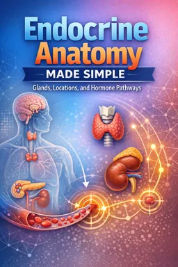
Example you can visualize anatomically: thyroid hormones released from thyroid follicles enter thyroid capillaries, drain into thyroid veins, return to the heart, and then reach many tissues (e.g., skeletal muscle, liver) via systemic arteries.
Paracrine signaling (local diffusion in tissue)
Definition: chemical messenger diffuses through interstitial space to nearby cells within the same tissue region, without relying on long-distance bloodstream transport as the primary route.
Anatomy-based example: within the pancreas, islet cells can influence neighboring islet cells and nearby acinar tissue through local diffusion across short distances in the pancreatic interstitium. The message may still eventually enter blood, but the defining feature is local neighborhood action.
- Listen to the audio with the screen off.
- Earn a certificate upon completion.
- Over 5000 courses for you to explore!

Download the app
Autocrine signaling (self-targeting within the same cell population)
Definition: a cell releases a messenger that binds receptors on the same cell or the same cell type nearby, shaping its own activity.
Anatomy-based example: endocrine cells within a gland can modulate their own secretion through feedback onto receptors on the secreting cells themselves (local microenvironment action rather than systemic delivery).
Quick comparison table
| Type | Main anatomical route | Distance | Key identifying feature |
|---|---|---|---|
| Endocrine | Capillary → veins → heart → arteries → target capillaries | Long | Bloodstream is the delivery system |
| Paracrine | Interstitial diffusion within a tissue region | Short | Neighbor-to-neighbor signaling |
| Autocrine | Interstitial diffusion back to same cell type | Very short | Self-regulation via own receptors |
3) How hormones “choose” targets: receptor distribution meets tissue anatomy
Hormones do not “aim” for a specific organ. They circulate widely, but only tissues with the appropriate receptors respond. Targeting is therefore determined by receptor distribution, which is an anatomical and cellular property of tissues.
A) Membrane receptors: surface binding and fast signaling
Where the receptor is: embedded in the cell membrane.
What this implies anatomically: the hormone must reach the interstitial fluid around the cell and bind to the outer surface. This is typical for water-soluble hormones that do not readily cross lipid membranes.
- Pathway emphasis: delivery to target capillaries and diffusion across the capillary wall into interstitial space is crucial.
- Tissue example: vascular smooth muscle cells with membrane receptors can respond quickly when the hormone reaches their surrounding interstitial fluid.
B) Intracellular receptors: entry into cells and longer-lasting effects
Where the receptor is: in the cytoplasm or nucleus.
What this implies anatomically: the hormone must reach the interstitial fluid, then enter the cell to bind its receptor. This is typical for lipid-soluble hormones.
- Pathway emphasis: systemic distribution is broad, but response is limited to tissues whose cells contain intracellular receptors.
- Tissue example: many tissues respond when their cells contain nuclear receptors; the same circulating hormone can have different effects depending on receptor type and local gene programs.
Receptor distribution as an “anatomical map”
Imagine each organ has a different “receptor density.” Even if hormone concentration is similar in arterial blood reaching multiple organs, the functional target is the tissue with the receptor presence (and sufficient receptor number) to trigger a meaningful response.
4) Structured pathway diagram (gland → blood → target tissue)
Use this diagram format to practice tracing any endocrine hormone. Replace the placeholders with a specific gland and target.
[Gland endocrine cells] → (interstitial fluid) → [Gland capillary bed] → [Venous drainage] → [Venae cavae] → [Right heart] → [Lungs] → [Left heart] → [Aorta] → [Arterial delivery] → [Target organ arterioles] → [Target capillaries] → (interstitial fluid) → [Receptor-binding on/in target cells] → [Physiologic response]Diagram with “checkpoints” you should be able to label
- Checkpoint 1: Where does the hormone enter blood? (capillary bed of the gland)
- Checkpoint 2: Which vessels carry it away first? (venules/veins = venous drainage)
- Checkpoint 3: Which pump redistributes it? (left heart into systemic arteries)
- Checkpoint 4: Where does it leave blood? (target capillaries)
- Checkpoint 5: What makes the tissue a “target”? (receptor presence and type)
5) Practical step-by-step tracing examples (anatomy-first)
Example A: A thyroid hormone traveling to skeletal muscle
- Release: endocrine cells in the thyroid release hormone into local interstitial fluid.
- Entry: hormone enters thyroid capillaries.
- Drain: blood exits via thyroid venous drainage into larger veins.
- Return: venous blood reaches the right heart, passes through lungs, then to the left heart.
- Distribute: left heart pumps hormone-containing blood into the aorta and systemic arteries.
- Deliver: arteries to skeletal muscle deliver blood to muscle arterioles and capillaries.
- Exit: hormone moves from capillaries into muscle interstitial fluid.
- Bind: hormone binds receptors in muscle cells (receptor location determines whether binding is membrane-level or intracellular).
Example B: A pancreatic islet hormone traveling to the liver
This example emphasizes that some targets are reached through a particularly direct vascular route.
- Release: islet cells release hormone into pancreatic interstitial fluid.
- Entry: hormone enters pancreatic capillaries.
- Drain: venous blood from the pancreas flows into veins that contribute to the portal route toward the liver.
- Deliver: liver sinusoids (specialized capillaries) expose hepatocytes to the hormone.
- Bind: hepatocyte receptors determine response (membrane vs intracellular receptor location shapes the signaling style).
6) Mini-exercises: trace the path on a simplified circulatory map
Use this simplified map for all exercises. Your job is to write the missing labels and arrows for each hormone scenario.
Simplified circulatory map (template to reuse) 1) Gland capillaries → 2) Gland veins → 3) Large veins → 4) Right heart → 5) Lungs → 6) Left heart → 7) Aorta → 8) Target arteries → 9) Target capillaries → 10) Receptors in target tissueExercise 1: Endocrine vs paracrine decision
Prompt: A messenger is released in a gland and affects cells in the same gland region within seconds, without needing to pass through the heart first. Is this endocrine, paracrine, or autocrine? Write the route using either (a) interstitial diffusion only, or (b) the full circulatory template above.
- Your output format:
Type = ___; Route = ___
Exercise 2: Trace a “classic endocrine” route
Prompt: Choose any endocrine gland you know and a distant target (e.g., gland in the neck → target in the thigh). Fill in each of the 10 steps in the simplified circulatory map with the correct anatomical terms (capillaries, veins, heart, arteries, target capillaries, receptors).
- Your output format:
1) ___ → 2) ___ → ... → 10) ___
Exercise 3: Receptor-based targeting
Prompt: Two tissues receive the same hormone concentration in arterial blood. Tissue A has abundant receptors; Tissue B has none. Predict which tissue responds and identify the anatomical “decision point.”
- Your output format:
Responding tissue = ___; Decision point = ___ (capillary delivery vs receptor presence)
Exercise 4: Membrane vs intracellular receptor consequences
Prompt: For a hormone that binds membrane receptors, list the last two anatomical steps immediately before signaling begins. For a hormone that binds intracellular receptors, add the extra step required after leaving the capillary.
- Your output format:
Membrane receptor: ___ → ___; Intracellular receptor: ___ → ___ → ___